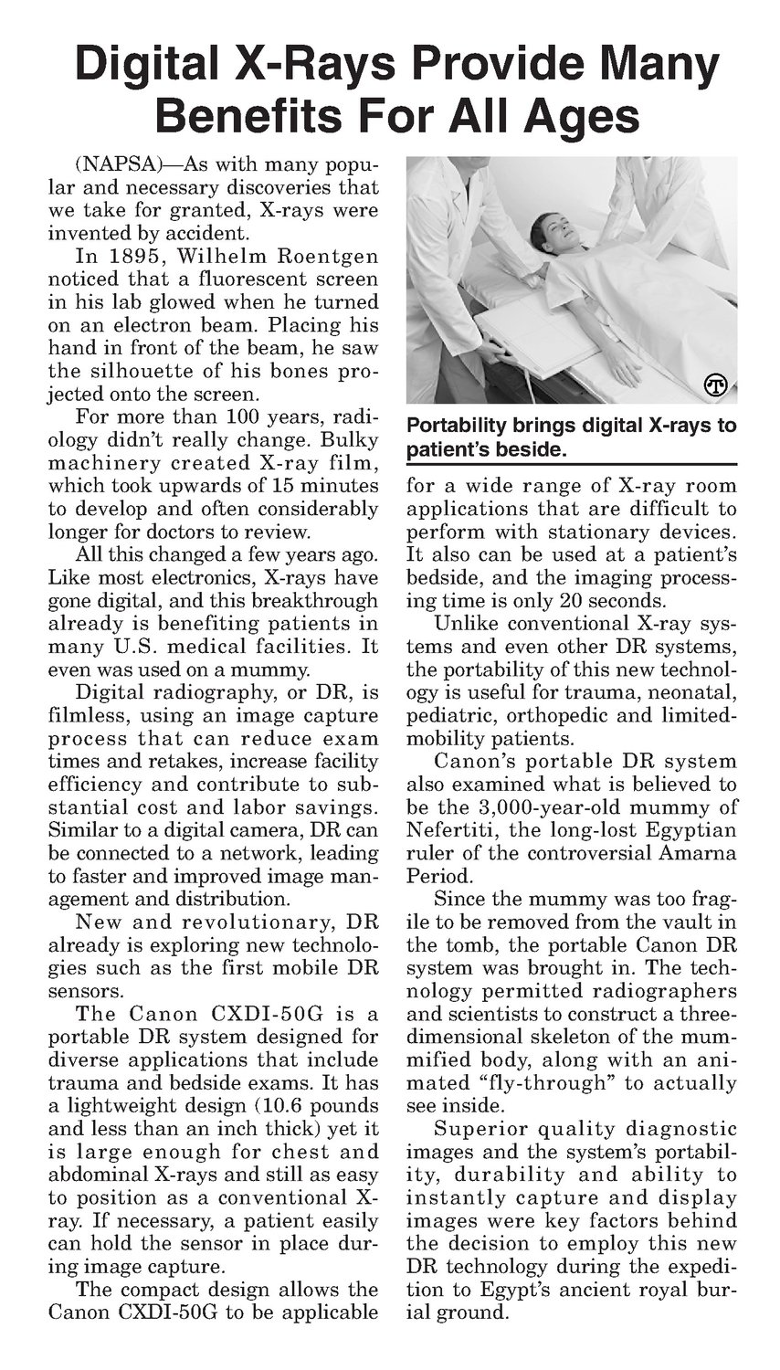
Benefits For All Ages (NAPSA)—Aswith many popular and necessary discoveries that we take for granted, X-rays were invented by accident. In 1895, Wilhelm Roentgen noticed that a fluorescent screen in his lab glowed when he turned on an electron beam. Placing his hand in front of the beam, he saw the silhouette of his bones projected onto the screen. For more than 100 years, radiology didn’t really change. Bulky machinery created X-ray film, which took upwards of 15 minutes to develop and often considerably longer for doctors to review. All this changed a few years ago. Like most electronics, X-rays have gone digital, and this breakthrough already is benefiting patients in many U.S. medical facilities. It even was used on a mummy. Digital radiography, or DR, is filmless, using an image capture process that can reduce exam times and retakes, increase facility efficiency and contribute to substantial cost and labor savings. Similarto a digital camera, DR can be connected to a network, leading to faster and improved image management anddistribution. New and revolutionary, DR already is exploring new technologies such as thefirst mobile DR sensors. The Canon CXDI-50G is a portable DR system designed for diverse applications that include trauma and bedside exams. It has a lightweight design (10.6 pounds and less than an inch thick) yet it is large enough for chest and abdominal X-rays andstill as easy to position as a conventional X- ray. If necessary, a patient easily can hold the sensor in place during image capture. The compact design allows the Canon CXDI-50G to be applicable Portability brings digital X-rays to patient’s beside. for a wide range of X-ray room applications that are difficult to perform with stationary devices. It also can be used at a patient’s bedside, and the imaging processing timeis only 20 seconds. Unlike conventional X-ray systems and even other DR systems, the portability of this new technology is useful for trauma, neonatal, pediatric, orthopedic and limitedmobility patients. Canon’s portable DR system also examined whatis believed to be the 3,000-year-old mummyof Nefertiti, the long-lost Egyptian ruler of the controversial Amarna Period. Since the mummywastoofragile to be removed from the vault in the tomb, the portable Canon DR system was brought in. The technology permitted radiographers and scientists to construct a threedimensional skeleton of the mummified body, along with an animated “fly-through” to actually see inside. Superior quality diagnostic images and the system’s portability, durability and ability to instantly capture and display images were key factors behind the decision to employ this new DR technology during the expedition to Egypt’s ancient royal burial ground.